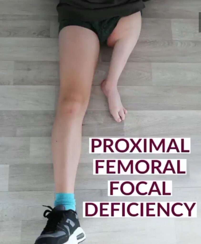
Proximal Femoral Focal Deficiency (PFFD) is a rare congenital condition affecting the femur, the bone in the thigh. Children with PFFD are born with an underdeveloped or deformed femur, which can vary in severity. This condition impacts a child\’s leg length, mobility, and overall development. In this blog, we’ll explore the causes and diagnosis of PFFD to help parents understand this complex condition better.
Causes of PFFD
The exact cause of PFFD remains unclear, but it’s believed to occur early in pregnancy, typically around the sixth week of fetal development. Various factors could contribute, such as:
1. Genetic Factors: Although PFFD is usually not inherited, in some cases, genetic mutations or disruptions during fetal growth may play a role.
2.Environmental Influences: Exposure to certain toxins, medications, or radiation during pregnancy may interfere with normal bone development in the fetus.
3.Vascular Issues: Reduced blood supply to the developing limb during pregnancy may hinder femur growth, leading to the characteristic deformities of PFFD.
While the exact cause is unknown, understanding these possible contributing factors can provide insight into how the condition develops.
Diagnosing PFFD
Diagnosing PFFD involves a combination of clinical examination and imaging studies. Early detection is crucial for determining the best treatment path and improving long-term outcomes for the child.
1. Physical Examination
At birth, doctors will often notice differences in the length and appearance of the legs. Children with PFFD may present with shorter legs, hip or knee deformities, and reduced range of motion in the affected limb.
2. X-Rays and Imaging
X-rays are typically the first step in diagnosing PFFD, allowing doctors to assess the severity of the femur abnormality. In some cases, advanced imaging like MRI or CT scans may be needed to examine the hip joint and other related structures.
3. Classification of PFFD
PFFD is categorized based on the severity of the femoral deficiency. These classifications help in determining the most appropriate treatment plan:
- Type A: Mild shortening with a nearly normal hip joint.
- Type B: Shortened femur with some hip joint deformities.
- Type C: Severe shortening with significant deformity of the hip and thigh.
- Type D: Complete absence of the femur.
This classification aids in developing a personalized treatment plan tailored to the specific needs of the child.
Why Early Diagnosis Matters:
Early diagnosis of PFFD is crucial for effective management, enabling timely treatments like limb lengthening, corrective surgery, or prosthetics, which improve mobility, quality of life, and long-term outcomes.
Conclusion
Proximal Femoral Focal Deficiency is a complex condition, but with proper diagnosis and early treatment, children can lead fulfilling and active lives. Understanding the causes and diagnostic process is the first step toward addressing the condition effectively. Dr. Sameer Desai, a leading pediatric orthopedic surgeon in Pune, is dedicated to helping families navigate the journey of PFFD treatment, ensuring the best possible care for your child.
If you suspect your child may have PFFD or want expert advice on pediatric orthopedic conditions, Dr. Desai is here to help.